Looking for design inspiration?   Browse our curated collections!

Previous Image

|

Next Image
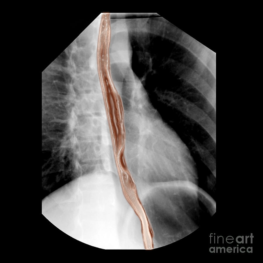
Diagnostic Imaging Photograph - Normal Esophagus #1 by Living Art Enterprises

Normal Esophagus #1 is a photograph by Living Art Enterprises which was uploaded on May 13th, 2014.

More from This Artist

View Profile

Fiber Tracts Of The Brain, Dti #4 by Living Art Enterprises
Carotid Duplex Ultrasound Exam #4 by Living Art Enterprises LLC
Cross-sectional Mri Of The Human Brain by Medical Body Scans
Roentgens X-ray Machine, 19th Century by Science Source
Coronal View Mri Of Normal Brain by Medical Body Scans
Chiari I Malformation MRI by Living Art Enterprises and Photo Researchers
Corona Radiata, Diffuse Tensor Imaging #1 by Living Art Enterprises
Pet Scan Of 20-year-old Brain by Science Source
Rotator Cuff Tear #2 by Medical Body Scans
Degenerative Disc Disease, Mri by Science Source
Normal Brain Diffusion Tractography by Living Art Enterprises
Normal Brain Diffusion Tractography #1 by Living Art Enterprises
Pierced Navel, X-ray by Scott Camazine
Optic Nerve Head Druzen #4 by Paul Whitten
Dural Arterial Venous Fistula, Angiogram by Living Art Enterprises
Atherosclerosis, Ct Angiogram #2 by Living Art Enterprises
Enhanced Composite Imaging Of Modern Man by Living Art Enterprises
Cerebral Angiogram #2 by Medical Body Scans
Chest X-ray by Ted Kinsman
Fiber Tracts Of The Brain, Dti #3 by Living Art Enterprises

Comments

There are no comments for Normal Esophagus #1.   Click here to post the first comment.

Recently Viewed

Normal Esophagus #1 by Living Art Enterprises
Previous Image Next Image